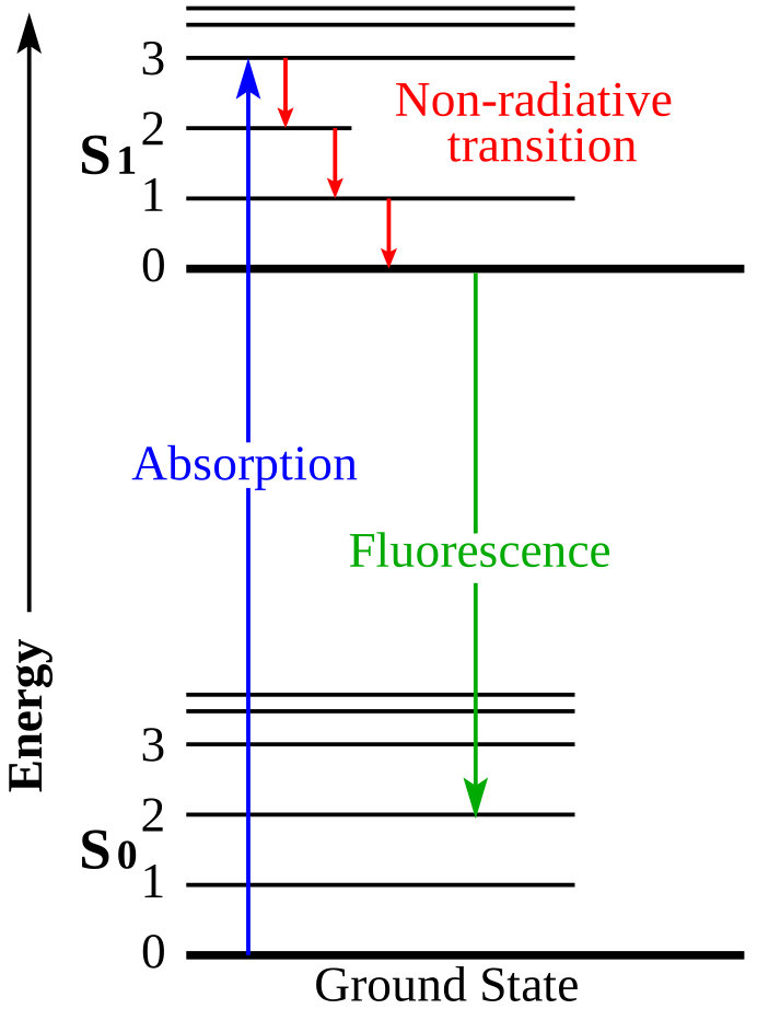
Energy levels, not anatomy. Blue: absorption. Red: non-radiative relaxation. Green: fluorescence. Original by Jacobkhed; vectorisation by Д.Ильин. Source · CC0 1.0.
Excitation
A photon supplies energy
Relaxation
Some energy is lost without light
Emission
A lower-energy photon is emitted
THE PRINCIPLE
How it works
A fluorescent agent absorbs light and emits light at a longer wavelength. The imaging system separates the returning fluorescence from illumination, detects it and presents a view the surgeon can interpret. For ICG, this uses the near-infrared part of the spectrum.
01
Read the energy-level drawing
Absorption raises the molecule to an excited state. After non-radiative relaxation, fluorescence returns it towards a lower electronic state. The emitted photon typically has a longer wavelength than the excitation photon.
02
Make the signal visible
The camera detects fluorescence outside the visible band and converts it to a display image. The assigned green or other overlay colour is a visual aid, not the literal colour of near-infrared light.
03
Keep the biological question separate
The mechanism explains how light is generated and detected. It does not, by itself, establish a clinical indication, diagnostic accuracy or benefit for a particular patient.
Energy levels, not anatomy. Blue: absorption. Red: non-radiative relaxation. Green: fluorescence. Original by Jacobkhed; vectorisation by Д.Ильин. Source · CC0 1.0.
NIR / ICG
A signal beyond visible light.
Near-infrared describes a wavelength range. ICG is a fluorescent agent that can be excited and detected within that range. Depending on the authorised application, it can support visualisation of perfusion or anatomical structures.
NIR excitation
ICG fluorescence
Filtered camera
Display overlay
Protein-bound ICG has reference absorption and emission peaks near 805 and 830 nm. These are not universal camera or LED specifications.
In blue-light cystoscopy, hexaminolevulinate supports the formation of fluorescent porphyrins in tissue. With suitable blue-light illumination and filters, fluorescence appears red against the blue background.
Blue excitation
Porphyrin fluorescence
Filtered view
White-light review
This is used alongside—not instead of—white-light examination. False signals and missed lesions remain possible. Other blue-light applications require their own agent and system assessment.
CYTALUX is a folate-receptor-targeted optical imaging agent, not a light colour or camera mode. A compatible NIR system detects its fluorescence during specified surgical applications.
Targeted agent
NIR excitation
Compatible detector
Surgeon interpretation
Its US label covers adjunctive identification of malignant ovarian lesions and malignant or non-malignant pulmonary lesions in adults with known or suspected lung cancer. Fluorescence alone does not establish malignancy.
These are different agent–illumination–detector combinations, not interchangeable features. Spectra’s ICG capability does not establish support for blue-light agents or CYTALUX. Confirm the exact device, agent, intended use and local approvals. Fluorescence supplements clinical assessment; it does not replace it.
 UNIVLABS