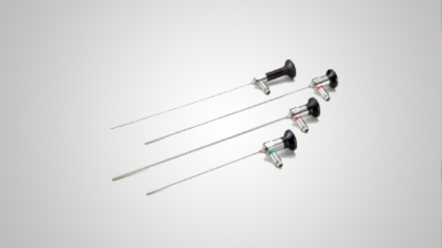
An endoscope brings illumination to the field and carries the image back to the camera or an integrated sensor. The scope, coupler, camera, light source and monitor work as one imaging chain. A high-resolution display cannot restore detail that was not captured.
01
Separate illumination from imaging
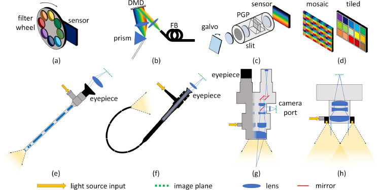
In a rigid optical laparoscope, illumination fibres carry light towards the operative field. An objective and relay-lens system convey the returning image to the eyepiece and camera. These are different paths within the same instrument.
02
Know where the sensor sits
A rod-lens telescope relays an optical image to an external camera. A chip-on-tip videoscope places its image sensor distally. Olympus ENDOEYE is a manufacturer example of the latter, not a specification for UnivLabs scopes.
03
Choose geometry for the procedure
Diameter, working length and direction of view are separate specifications. A 30° direction of view describes an oblique view; it does not mean a 30° total field of view. Verify the exact model and its NIR compatibility.
Read panel (e): the rigid laparoscope. Separate illumination and image paths are shown alongside other spectral-imaging configurations. Clancy, Jones, Maier-Hein, Elson & Stoyanov, Surgical spectral imaging (2020), Figure 2. CC BY 4.0.
 UNIVLABS